





Pinned Tweet
… and eventually like this!
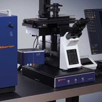
Indeed, it’s a building-brick model of our MIRAVA POLYSCOPE – limited edition! 🤩
Want to have one? Then sign up to our newsletter: link.abberior.rocks/newslett…
Each quarter, we raffle 1 set among all new subscribers!
#MIRAVAPOLYSCOPE

🏆STEDYCON 2 has received the 2026 Microscopy Today Innovation Award!
STEDYCON 2 was recognized as “the first modular add-on system combining multicolor STED and phasor-based FLIM (TIMEBOW) in an alignment-free, browser-controlled platform.”
link.abberior.rocks/stedycon


✨ FLIM is now available for STEDYCON!
Experience fluorescence lifetime imaging with the same ease of use and intuitive operation you know from STEDYCON!
STEDYCON is your fast track to lifetime imaging!
link.abberior.rocks/timebow

This week the first #MINFLUX image in Japan has been successfully acquired using our MIRAVA POLYSCOPE at CiDER, Osaka University.
We are excited to bring cutting-edge super-resolution microscopy technology to researchers around the world. 🌍
Learn more: abberior.rocks/mirava-polysc…


🔬 Meet STEDYCON 2 – STED imaging has never been easier (or more fun)!
Discover what STEDYCON 2 has to offer:
👉 Confocal & STED microscopy
👉 TIMEBOW fluorescence lifetime imaging
👉 Flexibility, reliability & stability for your experiments
link.abberior.rocks/stedycon

Organelle architecture in 3D: nuclear pore complexes and Golgi visualized throughout a full spheroid with abberior #STARRED and #STARORANGE, actin with #STARGREEN Phalloidin, nuclei with DAPI.
NIH-3T3 spheroids, imaged on the abberior #MIRAVA Polyscope with ibidi µ-Slides.
This week, we celebrated the opening of a new imaging system at the Third Faculty of Medicine at Charles University in Prague.
The installation combines our #MIRAVAPOLYSCOPE with a #CrestOptics spinning-disk system on a single microscope platform.
Thanks to everyone involved!


Confocal or STED? Why not both? 🔬
STED builds on confocal microscopy to reveal details beyond the diffraction limit.
Learn when to use each and why they complement each other:
abberior.rocks/knowledge-bas…


MIRAVA rocks at #SPAOM2026🔬✨
We’re excited to be at SPAOM in Vic (Barcelona), from October 6–9 with our MIRAVA.
Visit our booth, meet the team, and discover MIRAVA live in action. We’re looking forward to great conversations, new connections, and super-resolution microscopy!

Cardiomyocytes are a key model system for studying heart disease. LAMP1 (yellow, STAR RED), TOM20 (cyan, STAR ORANGE), Myosin (red, STAR GREEN). Imaged at Fraunhofer ITMP Göttingen by Fabian Link. #Cardiomyocytes #STARdyes #abberior #FluorescenceFriday

Mitochondria meet lysosomes 🔬
Fibroblasts: Mic60 (STAR RED), LAMP1 (STAR ORANGE), DAPI
Imaged by Fabian Link at Fraunhofer ITMP Göttingen.
#Microscopy #CellBiology #STARdyes

Ever wanted to build your own microscope? 🔬
How about starting with our MIRAVA POLYSCOPE Building Brick Model? 🤩
Today is your last chance to enter our Q3 raffle! Sign up for our newsletter: link.abberior.rocks/newslett…

Last week we trained the new #MIRAVAPOLYSCOPE at Crick Advanced Light Microscopy (CALM) Facility in London together with PHOTON LINES🔬.
It was great to explore and train the system together with Rocco D'Antuono and Rosalind Norkett.
Happy Imaging with the new MIRAVA!


Nuclear pore complexes stained for Mab414 with abberior STAR 680🟦, ELYS with abberior STAR RED🟥, and Nup96 with abberior STAR ORANGE🟨. STED resolves individual nuclear pore complexes in three colors. Image quality is enhanced using #TRUESHARP deconvolution.
#fluorescencefriday

A new chapter for #super-resolution #microscopy in #Hamburg: abberior Instruments and the UMIF Facility at #UKE are establishing a new Reference Center.
Join us for the opening on November 10, with talks from Nobel laureate Prof. Hell, Prof. Lechner & Prof. Meyer-Schwesinger.


“Deeper imaging” is not a single optical problem.
Choosing the right technique means understanding what degrades the image with depth—and when aberration correction is needed.
Learn more about confocal vs. multi-photon microscopy:
abberior.rocks/knowledge-bas…

Nuclear pore complexes labelled for NUP96🟥 and Mab414🟦, imaged from confocal to high-power STED.
Each imaging mode reveals progressively finer structural details.
One sample, multiple imaging modes - with the #MIRAVAPolyscope.
#fluorescencefriday

Why does confocal microscopy look clearer than wide-field imaging?
It’s not just resolution — it’s about rejecting out-of-focus fluorescence.
Learn how optical sectioning improves contrast and how array detection can reduce background even further:
abberior.rocks/knowledge-bas…
