

Daily research insights on whiplash, neck pain & MVA recovery. Evidence-based care that gets patients back to life. 🏥
Petaluma. CA
Joined February 2026
- Tweets328
- Following76
- Followers10
- Likes428





What Helps and What Slows Healing
Graded activity helps. Movement within what the tissue can handle, increased in steps as it recovers. Prolonged rest delays recovery, and loading healing tissue too much or too soon can stop repair.
What else helps: enough food and protein (one review suggests 2 to 2.5 grams per kilogram per day during immobilization), managing stress, and sleep.
What slows it: smoking (about twice the odds of delayed wound healing), uncontrolled diabetes, older age, alcohol, and the wrong dose of load.
Anti-inflammatory drugs (NSAIDs) are unclear — most of the research is in animals and cell cultures, and human studies are few. Medication decisions belong with your physician, so ask before starting or stopping anything.
4
Feeling better doesn't mean the tissue is done healing.
In athletes cleared to return to sport after a hamstring strain, 89% still showed abnormal signal on MRI. Remodeling runs for months in most soft tissues, so the weeks after pain fades are still a time to build activity back up gradually.
Pain can also last after the tissue has healed. After whiplash, people who still had moderate or severe symptoms at six months already showed widespread sensitivity within the first month — a sign of changes in how the nervous system processes pain.
But lasting pain often has a physical source too. In one study, the facet joints accounted for 60% of persistent neck pain after whiplash. Pain can have more than one cause, and it's real.
2
Why does a sprained ligament take months while a muscle strain heals in weeks?
Blood supply.
Healing needs oxygen, nutrients and cells. Blood delivers all of them. Muscle is well supplied. Ligaments and tendons use about 7.5 times less oxygen than muscle, and that low metabolic rate means slow healing.
The center of a spinal disc has no blood supply at any age. In adults, only the outermost layers keep blood vessels. That's why disc injuries heal the least of any tissue — they can't get what they need to rebuild.
Some tissues regenerate: bone rebuilds with new bone, and muscle regrows its own fibers. Ligaments, tendons and discs repair with scar. The scar works, but it never matches the original tissue's strength or structure.

3
Every injured tissue heals in three overlapping phases, whether it's muscle, ligament, tendon or disc.
First: inflammation in the first few days — white blood cells clear debris and set up repair.
Second: repair over the following weeks — quick type III collagen patches the gap.
Third: remodeling for months to years — that patch is replaced with stronger type I collagen that lines up with the loads it carries.
The phases are the same. The timeline for each tissue is not. Muscle, with rich blood supply, completes repair in 3 to 4 weeks. Ligaments take at least 6 weeks to 3 months, and they're still remodeling a year later.
Blood supply is the difference.
5
The radiologist wrote 'age-appropriate degeneration' on your report. Here's why that's misleading:
If this was about your age, we'd see the same changes at every level of your spine — because your whole spine is the same age.
But four of your five discs look completely normal. Only L4-5 shows the herniation.
Age doesn't pick and choose. The crash does.
You may have had some wear-and-tear before — most people do, and it causes no symptoms. But the crash turned silent degeneration into a symptomatic injury.
That's causation. The crash caused it."
Patients need to understand the rebuttal well enough to repeat it when questioned by adjusters or reviewers.
12
Weak: "MRI shows age-appropriate degenerative changes at L4-5 with herniation likely related to trauma."
This concedes the radiologist's framing and adds causation as an afterthought. Easily dismissed.
Strong: "MRI demonstrates focal disc herniation at L4-5. Review of all lumbar levels reveals normal disc morphology at L1-2, L2-3, L3-4, and L5-S1, indicating the L4-5 pathology is level-specific rather than age-related. The mechanism of injury directly explains the L4-5 herniation. Any characterization of this finding as 'age-appropriate degeneration' is contradicted by the normal appearance of adjacent levels in this same-aged patient."
Don't accept their framing. Challenge it with logic and imaging evidence.
12
Insurance companies conflate two very different concepts to deny claims:
Pre-existing condition: You were being treated for it the day before the crash.
Predisposing condition: It existed silently, caused no symptoms, required no treatment — but made you more vulnerable when trauma occurred.
A degenerative disc that was asymptomatic before a collision is NOT a pre-existing injury.
It's a predisposing factor.
And predisposition doesn't negate causation — it supports it. The collision is still what caused the disc to fail.
The legal doctrine is clear: you take your victim as you find them. Pre-existing vulnerability doesn't reduce liability. It may increase damages.
6
A 45-year-old patient's MRI shows one herniated disc at L4-5. The radiologist calls it "age-appropriate degeneration."
Here's the problem:
The L3-4 disc above it? Normal.
The L5-S1 disc below it? Normal.
All five lumbar discs are 45 years old. Same patient. Same age. Same genetics. Same time.
If the L4-5 herniation is truly caused by age, why don't the other four discs show the same pathology?
Age is systemic. It doesn't pick one disc and spare the rest.
Level-specific pathology requires a level-specific cause — and in a post-collision patient, that cause is the collision.

15
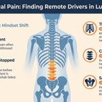
Patients with persistent lumbar pain who don't respond to correct local treatment have an unaddressed remote driver — until proven otherwise.
The regional interdependence literature gives three places to look:
→ The thoracic spine, for mobility restriction driving both cervical and lumbar compensation
→ The cervical spine, for proprioceptive deficit driving lumbar motor control failure
→ The sacroiliac joint, for pelvic asymmetry driving the entire posterior chain
The patient isn't failing rehab. The assessment stopped one region too early.
Expand the examination before concluding the diagnosis is wrong or the patient is non-compliant.

22
Your cervical spine's proprioceptors don't just report head position — they coordinate trunk motor control throughout the spine.
Hodges and Richardson documented that the deep spinal stabilizers (multifidus, transversus abdominis) activate before a limb moves, protecting the spine from the incoming load.
This feedforward activation depends on cervical proprioceptive input.
When cervical proprioception is disrupted — by joint capsule injury, deep muscle dysfunction, or post-collision sensorimotor deficit — the feedforward activation delays or fails.
The lumbar spine absorbs the unprotected load. Repeated unprotected loading produces lumbar pathology.
The neck injury happened first. The back pain came second. Most patients never hear that the two are connected.

8
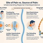
Treating the site of pain is not the same as treating the source of pain.
A patient with low back pain from lumbar hyperlordosis — driven by a forward head posture caused by cervical extension restriction — will cycle through lumbar treatment indefinitely.
The lumbar mechanics improve during treatment. The cervical restriction reloads the lumbar segment the moment rehabilitation ends.
The symptom is real. The diagnosis is accurate. The treatment region is wrong.
Regional interdependence means the spine has one geography of pain and another geography of cause. Clinical assessment that stops at the site of the complaint misses the second map entirely.

10
Atlas retweeted

A car may run over a small object with limited damage, but a motorcycle may lose stability, slide, fishtail, throw the rider, or force an emergency swerve into another lane.
Learn More: avrek.com/blog/los-angeles-m…

Thoracic spine manipulation reduces cervical pain and improves cervical range of motion in patients who never had a thoracic complaint.
That finding — documented by Cleland and colleagues in a randomized clinical trial — is inexplicable if the spine works as isolated regional segments.
It only makes sense if the spine works as a system.
What this means in practice: if your cervical treatment is technically correct and producing incomplete results, the thoracic spine is the first place to look.
Not because the cervical diagnosis is wrong — but because the cervical segment is absorbing compensation for a restriction that lives two regions away.
28
Who recovers from whiplash and who doesn't is predicted far less by imaging than by the proprioceptive deficit at initial evaluation.
Patients with high initial joint position error — poor accuracy when repositioning the head with eyes closed — are more likely to develop chronic symptoms. Not because the mechanical injury is worse. Because the sensorimotor deficit is deeper, and if it isn't specifically targeted in rehabilitation, it doesn't resolve on its own.
Strength training doesn't close that gap. Sensorimotor retraining does. The patients who recover fastest are the ones whose rehabilitation starts at the nervous system and works outward — not the ones who wait for pain to subside before they begin.
7
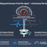
Within the intervertebral foramen of your cervical spine, the facet joint capsule contains mechanoreceptors that continuously report head position to the brain. When the cccapsule is strained in a rear-end collision, those receptors are disrupted. The brain loses accurate position data.
To manage the uncertainty, it raises its threat estimate. It outputs more pain. This is not damage-in-progress — it is a recalibrated threat response to imprecise sensory information.
The input the brain needs to revise that response is movement: controlled, precise, proprioceptively rich movement that restores the signal the disrupted capsule can no longer provide reliably.
This is why sensorimotor retraining reduces pain. Not by repairing tissue. By restoring information.

7
Rest after a whiplash injury does not protect your spine. It trains your nervous system to stay in pain.
When the neck stops moving, the large-diameter sensory fibers that close the pain gate go silent. Nociceptive signals flow with less inhibition. Pain increases — not because the injury is worse, but because the brake on pain transmission has been switched off by immobility.
Cervical collars produce worse outcomes than early active mobilization across multiple randomized controlled trials. The thing that feels protective is making recovery slower. The thing that feels dangerous — controlled, guided movement — is what interrupts the pain cycle.
3
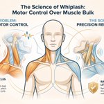
The deep cervical flexors — the muscles that actually stabilize your cervical spine — reduce their activation after whiplash injury. Not because they're weak. Because the sensory disruption from the trauma changes the motor control pattern.
The superficial muscles compensate. They load structures that are already sensitized. They perpetuate the pain cycle. The fix isn't to strengthen the superficial muscles — they're already overactive. The fix is to retrain the deep system through precision movement and sensory feedback.
Jull and colleagues demonstrated this with Level 1 evidence in a randomized controlled trial. The injury disrupts motor control. Rehabilitation has to target motor control — not just muscle bulk.

4
Across 29 studies examining predictors of chronic disability after whiplash, imaging findings were not a significant predictor of who recovered and who didn't.
The factors that predicted chronicity: high initial pain intensity, restricted cervical range of motion, neurological symptoms at presentation, and elevated psychological distress. Not the scan. This is from Scholten-Peeters et al., 2003 — a systematic review available for over twenty years.
What this means in practice: the patient whose scan looks normal but who rates their pain 8/10, can't turn their head past 30 degrees, and hasn't slept a full night in six weeks is statistically at higher risk for chronic disability than the patient with a visible disc bulge and manageable pain. Function predicts the trajectory. Film doesn't.
The right question after a collision isn't "what does the scan show?" It's "what can this person still do — and what can't they do anymore?"
4
Your arm is going numb after a car accident. Your MRI is normal — no disc herniation, no spinal cord compression. Here's what's actually happening.
The dorsal root ganglion (DRG) — the cluster of sensory neuron cell bodies that sits in your intervertebral foramen — has one of the poorest blood-nerve barriers in the nervous system.
When adjacent capsular ligaments are torn in a collision, they release inflammatory mediators that diffuse directly to the DRG. The ganglion becomes chemically sensitized. It fires in dermatomal patterns. Your arm hurts and goes numb in exactly the way it would if a disc were compressing the root — because the physiological process is the same. Only the cause is different.
This is called chemical radiculitis. It produces symptoms indistinguishable from structural compression. It doesn't appear on MRI. EMG is typically normal because conduction isn't blocked — only sensitivity is altered. The nerve is irritated. The imaging is clear. Both things are true at the same time.

13
A normal scan after a car accident is not evidence that no injury occurred. It's evidence that no bony injury occurred — which is exactly what X-ray was designed to detect.
The injuries most common in rear-end collisions — partial ligament tears, capsular disruption at the facet joints, intramuscular microtears, dorsal root ganglion irritation — are systematically below the detection threshold of standard clinical imaging. This isn't a diagnostic gap that better technology has closed. It's a fundamental mismatch between the tool and the tissue being evaluated.
Insurance adjusters use "negative radiographic evidence" to deny claims. The problem is that negative radiographic evidence, in a low-speed rear-end collision, tells you almost nothing about whether soft tissue injury occurred. The research literature on this has been clear for over two decades. The claims-management practice hasn't caught up.
6