

An #OpenAccess science journal published by the Feinstein Institutes for Medical Research and @BioMedCentral @SpringerNature. Tweets curated by senior editors.
- Tweets290
- Following1.5K
- Followers696
- Likes2K


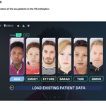


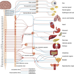



ALT Appendix E Visual illustration of the six patients in the VR orthoptics simulator


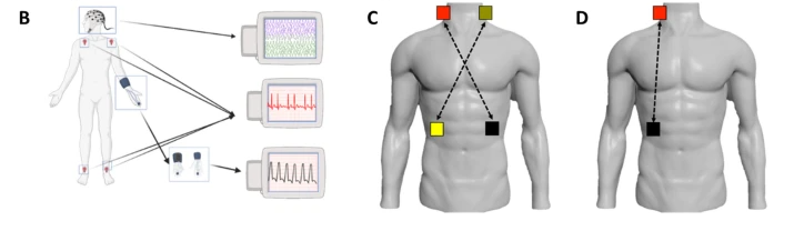
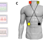
ALT Fig. 1 B- Physiological monitoring. Signals for each participant were recorded by a six-lead electrocardiogram (in red, wires attached to four foam adhesive electrodes placed at each shoulder and each ankle) and noninvasive blood pressure (in blue, small inflatable cuff on middle phalanx of middle finger). A dry electroencephalography system was placed on the participant’s head, while eye tracking glasses were used to record pupil diameter and gaze location. C-Bilateral stimulation montage. Adhesive electrodes were placed on each side of the neck, between the ear and jawline, and each side of the torso, below the ribcage on the midclavicular line. The return for each neck electrode was on the contralateral torso electrode. D One-sided stimulation montage. Adhesive electrodes were placed on one side of the neck, between the ear and jawline, and the return electrode was on the ipsilateral side of the torso, below the ribcage on the midclavicular line. Shown is the right side only


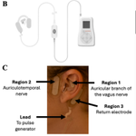
ALT Fig 1. B- Transcutaneous auricular VNS (taVNS) of auricular branch of the vagus nerve (ABVN) or tAN (stimulation of auriculotemporal nerve (ATN) and ABVN) administered via Spark Volta device. C- Electrode positioning for taVNS (regions 1 and 3) or tAN (regions 1, 2 and 3). Region 3 is the return electrode. Adapted from Czura et al. (Czura et al. 2026)


