

Become a better endoscopist with EndoCollab, the world's most comprehensive endoscopy community. Join us: https://xitter.cf/t.co/EkRtunpQyB
🌎
Joined February 2021
- Tweets10.2K
- Following2.9K
- Followers31.2K
- Likes21.2K


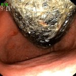


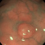

Small bowel bleeding, 8 mistakes:
1. DBE before repeat EGD/ileocolonoscopy
2. Trusting a negative capsule
3. No CTA if massive, no CT tumor
4. Overt: up to 67% in 48h, 18% day 5
5. Anatomy
6. Anastomosis, LVAD
7. No biopsy
8. APC at 90 or on clips
Which do you see most?

Full lecture on EndoCollab: endocollab.com/blogs/full-co…


EndoCollab™ | GI Endoscopy Community retweeted

Huge ulcer. Big vessel.
Hematemesis and hemorrhagic shock.
A one-stop hemostatic procedure was accomplished with an 11/6t over-the-scope clip.
The OTSC nicely compressed the ulcer bed and the massive visible vessel.
Case by Klaus Mönkemüller, MD, PhD.


Vessel plus Surface (VS) Classification (Yao Classification).
Microvascular (V) pattern: regular, irregular or absent.
Surface (S) pattern: regular, irregular or absent.


Two stents, one wire.
The pusher pushes both stents, one after the other.
The speaker has used this in traditional ERCP, in ERCP through a colonoscope, and here during double-balloon ERCP.
Have you placed two stents over one wire?
What makes a colon polyp difficult?
Shape: flat, carpet-like, depressed center.
Size: over 3 cm, or diminutive.
Site: right colon, behind folds.
Histology: serrated or dysplastic.
Prior biopsy, tattoo, or resection.
From Klaus Mönkemüller's Complex Colon Polyps lecture.

Klaus teaches this at 1:58 in his new 66-minute atlas, The Complex Colon Polyp (in the free preview): endocollab.com/blogs/full-co…

Massive gastric body and fundus folds on endoscopy.
What is your top diagnosis, and which biopsies do you take?
Reply below.

Papilla morphology by type: Type 1 (regular), Type 2 (small), Type 3 (protruding) and Type 4 (creased/ridged).
For each type the table gives prevalence, cannulation failure (OR, with Type 1 as reference), difficult cannulation and pancreatitis.


EndoCollab™ | GI Endoscopy Community retweeted
Common misconception: APC only causes superficial damage.
Per the authors, persistent APC on one spot can cause carbonization, deep tissue injury, even perforation. Apply it dynamically.
By Adil Mir, MD, Reid Wasserman, DO and Klaus Mönkemüller, MD, PhD
EndoCollab Quiz
White mucosal changes in the duodenum.
What is this?
a. Lymphangiectasia
b. Adenoma
c. Cholesterol plaques
d. Lipid deposits
Vote in the reply to see the answer.
Bonus: Including a free flash card

Answer in our Telegram quiz. Vote to see it:
t.me/endocollab/27

EndoCollab™ | GI Endoscopy Community retweeted
Ischemic colitis: rectal sparing, segmental left colon disease, and a linear ulcer on the mesenteric border (Zuckerman's sign). Case by Klaus Mönkemüller, MD, PhD.


How to Use Clips for Hemostasis:
> Anatomy and Mechanism of Operation of Clips
> Are They Single Use, Do They Rotate, How Much Tissue Can They Grasp?
> There is a Large Variety of Through-The-Scope Clips
> Technique of Clip Placement


