

A quantitative cell biologist who studies how force-generating cytoskeletal systems organize biological function across scales. Married to @gillianhoo.
Nashville, TN
Joined January 2015
- Tweets3.3K
- Following1.3K
- Followers33.2K
- Likes2.1K




Pinned Tweet
I am excited to finally be able to share with you our work reporting the first Extracellular Vesicle with a personality! This video does not show a cell, it is a Blebbisome! #CellBiology
nature.com/articles/s41556-0…
This cell photographed through a microscope kind of looks like a person. DNA in the nucleus and mitochondria are shown. #CellBiology


Another high profile paper showing the formation of blebbisomes from another cell type and not even a citation of the blebbisome paper...... Really???? My talk at AAEV next month is going to be fun for me........... aaev.org/
Dylan Burnette retweeted

Graduate student @EmmaKoory in Dylan Burnette's lab (@MAG2ART) received an honorable mention in the 2026 Nikon Small World competition for a video of a heart cell forming sarcomeres that drive contractions. @VanderbiltCBD
When “Better” Microscopy Becomes Fiction
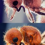
In light of the current controversy surrounding the use of AI in the Nikon Small World in Motion competition, I want to point out that AI enhancement of still microscopy images may be even harder to detect. With a movie, our eyes have temporal information that can sometimes clue us in to the AI-ness of what we are seeing; with a single image, that information is gone. Here is an example. The top image is the best reflected-light image I was able to acquire when I was young and had time to do such things. I never had the proper equipment for this type of imaging, which is essentially macrophotography through a microscope. I was simply holding a cheap digital camera up to the eyepiece by hand, and this was the best result I could get. The image below was generated by giving that original image to ChatGPT with the simple prompt, “Make this image of an ant that I acquired using a microscope better.”
The original image contains limitations: blown highlights, relatively low local contrast, noise and grain, limited depth of field, and regions where morphology simply isn’t resolved. The AI version doesn’t merely suppress those imperfections; it converts uncertainty into apparent information. The head acquires extremely convincing cuticular texture, the compound eye gains a beautifully resolved ommatidial lattice, individual hairs become sharply defined, thoracic surface structures become crisp, and the mandibles and antennae become more cool looking. Importantly, these additions are biologically plausible. Nothing immediately screams “AI.” That is what makes this fundamentally different from ordinary sharpening, denoising, deconvolution, or contrast adjustment. The resulting image invites the viewer to make biological observations about structures that may never have been recorded by the camera in the first place.

This took him two tries. For reasons that I do not understand, the first time Zach submitted this movie, it was not picked by the Small World judges. Its also too bad we did not publish this line of work. We all got caught up with the blebbisomes :-)

En este increíble video podemos observar cardiomiocitos, las células especializadas que impulsan las contracciones rítmicas del corazón, es decir, una coreografía microscópica. 🫀🔬
Los destellos azules representan ondas de calcio, que es la forma en que estas células se comunican y sincronizan los latidos. Las formas naranjas, son mitocondrias que suministran la energía necesaria para cada latido, garantizando que el corazón cuente con la energía requerida para contraerse de manera eficiente y continua.
©️ Dr. Zachary Sanchez, Nikon Small World in Motion.
youtube.com/watch?v=YTdWw09c…
There are a few things I love about this movie of a cell taken through a microscope.
This is the nucleus and perinuclear region of a living cell, imaged using two very different microscopy techniques: differential interference contrast (DIC) microscopy on the top and spinning-disk confocal fluorescence microscopy on the bottom.
The cell is expressing GFP, but in this experiment, the GFP isn’t attached to anything. We’re basically letting it go wherever it can go.
And that turns out to be pretty fun.
Free GFP fills much of the cytoplasm and nucleus, but there are some conspicuous places where it is largely absent. One is the nucleolus, the two large darker-looking region within the nucleus. The nucleolus is a specialized, densely organized compartment where ribosomal RNA is produced and the early stages of ribosome assembly take place.
GFP is also excluded from the interiors of membrane-bound organelles such as lysosomes and endocytic vesicles. In this movie, I can’t tell you which individual vesicles are lysosomes versus other endocytic compartments just from their appearance—but you can clearly see that there are lots of cellular structures that GFP does not enter.
And that gets to another reason I like this movie: it illustrates why we often use transmitted-light and fluorescence microscopy together.
Look at the DIC image on top. There is an enormous amount of cellular structure visible. DIC converts differences in optical path length into contrast, so we can see the nucleus, nucleolus, vesicles, boundaries, and all sorts of other structures without labeling any of them.
The catch is that we generally don’t know their molecular identity just from DIC.
Now look at the fluorescence image on the bottom. There is much less structural information, but what we see has molecular identity. We know that signal is GFP because GFP is what we are exciting and detecting.
That tradeoff is fundamental to how we image cells.
Transmitted-light techniques such as DIC can reveal a remarkably rich cellular landscape, but much of that landscape is molecularly anonymous. Fluorescence microscopy lets us ask where particular molecules are, but we can only label and distinguish a limited number of molecular species or cellular structures at once.
Put the two together, and you get something more useful than either alone: cellular context plus molecular identity.
Sometimes a simple movie is a nice reminder that how we choose to look at a cell determines what we are able to know about it.
Four cancer cells displaying continuous membrane blebbing videoed through a DIC microscope. No, these cells are not dying. #CellBiology
While we do not study apoptotic bodies in my lab, they are pretty cool! This cell expressing a membrane marker was videoed through a microscope while it was dying by @EmmaKoory. The video captured just the bottom of the cell. The cell rounds up as dying cells tend to do (i.e., it disappears from the region being imaged). Then apoptotic bodies come down and do cool stuff! In case you were wondering...... Yes, I am known internationally for my use of precise scientific language like "cool stuff" :-) #CellBiology
Cellular blebbiing and blebbisome formation are indeed different. Blebbisomes form from a large retraction fiber being detached from a migrating cell. It can be confusing as I came up with the named "blebbisome" because they often have membrane blebs themselves.
In contrast, It is thought that a membrane bleb can also detach from a cell to create another large extracellular vesicle called a "microvesicle". This type of extracellular vesicle has been given multiple names over the years.
Whether or not a blebbisome can form its own microvesicles has not been published. Spoiler alert: it can. #CellBiology

Four cancer cells displaying continuous membrane blebbing videoed through a DIC microscope. No, these cells are not dying. #CellBiology
A cell dividing into two new daughter cells photographed through a microscope. #CellBiology

Pictures of two insects acquired with microscopes hundreds of years apart. One was hand drawn and one was photographed using a digital camera. Can you tell which one is which?

A cell photographed through a microscope. DNA in the nucleus (purples), the Golgi apparatus (greens), and the actin filament cytoskeleton (oranges) are shown. #CellBiology

The mouth of a sand dollar photographed through a microscope. Not a mouth you say? OK. Peristomial aperture if you want to be fancy about it. #CellBiology

DNA in the nuclei of cancer cells videoed through a spinning disk confocal microscope. The first cell going through cell division got the assignment: one nucleus becomes two nuclei. The second cell, not so much. #CellBiology
A single cell photographed through a microscope. DNA in the nucleus (cyan), mitochondria (yellow), and actin filaments (magenta) are shown. #CellBiology

The powerhouses/overlords of the cell, mitochondria, photographed through a microscope. It’s just one cell. Well, just part of one cell—the perinuclear region. #CellBiology

Three cells photographed through a microscope. DNA in the nucleus (blue), mitochondria (green), and actin filaments (red) are shown. #CellBiology

A neuronal growth cone extending from a neuron taken from a sea slug (yes a sea slug is exactly what it sounds like) photographed through a microscope. Actin filaments and microtubules are shown. #CellBiology
