

Online-only Nature Research journal that publishes basic, translational, clinical and public health research in cardiac, vascular and blood biology.
- Tweets725
- Following936
- Followers7.8K
- Likes408
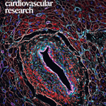








ALT Image: Yuka Morikawa at The Texas Heart Institute at Baylor College of Medicine. Cover design: Francesca Corra Microvascular senescence in single ventricle disease Image shows IL6 expression in samples from individuals with hypoplastic left heart syndrome. Red denotes IL6; purple denotes endothelial cells; green denotes cardiac fibroblasts; white denotes cell boundaries; and blue denotes nuclei.

ALT Image: Jun Chen Laboratory, University of California, Los Angeles. Cover design: Bethany Vukomanovic Magnetoelastic stent for restenosis diagnosis Chen et al. describe a smart stent that combines mechanical support with continuous blood-flow sensing and demonstrate its functionality in vivo for restenosis diagnosis.


ALT Image: Martin Dominguez, Drexel University, Irfan Kathiriya, University of California, San Francisco. Cover design: Bethany Vukomanovic TBX5 sets boundaries for a healthy heart Kathiriya et al. identify a Tbx5 + /Mef2cAHF + cardiac progenitor lineage that demarcates the interventricular septum during development, and changes in this lineage contribute to congenital heart defects in mice. (see Kathiriya et al.)


ALT Image: Bethany Vukomanovic. Cover design: Bethany Vukomanovic Genetic insights into long COVID Human genetic analyses show that thromboembolism-related variants are associated with an increased risk of long COVID, suggesting shared pathogenic pathways and implicating the thrombin receptor PAR-1 as a potential contributor. (see Schuermans et al.)


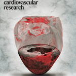
ALT Image: Leonardo Sacconi, Institute of Clinical Physiology. Cover design: Bethany Vukomanovic Cardiac fibrosis and arrhythmia Three-dimensional structural reconstruction of entire cleared mouse ventricles from a diseased heart acquired using mesoSPIM tomography. Myocardium is visualized by fluorescence (gray), and fibrosis is detected via scattered light (red) (see Giardini et al.).


ALT Image: ScienceBrush Design. Cover design: Bethany Vukomanovic; RNA editing in atherosclerosis: Weldy et al. show that expression of the RNA-editing enzyme ADAR1 in smooth muscle cells regulates activation of the double-stranded RNA sensor MDA5, in a new model of atherosclerosis.
