

Professor of Cell Biology at Universitat Politècnica de València, with a particular passion for molecular medicine and a strong interest in natural philosophy.
Valencia
Joined March 2025
- Tweets5.1K
- Following499
- Followers2.9K
- Likes5.4K




Pinned Tweet
Stellar Moments in Biology: A Journey Through the Discoveries That Explained Life.
A guide to the great questions of biology and to the remarkable moments when new evidence changed the way we understand life.
Definitively, the book I would have loved to have during my own university studies.
For a limited time, the ebook is available in your local amazon site for only US$1.50. The paperback edition is also available worldwide.
a.co/d/03159MWD

Rafael Sirera retweeted
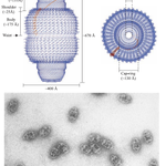
Surprisingly, vaults were only discovered in 1986, despite being considerably larger than ribosomes!
Vaults are large, barrel-shaped non-membranous ribonucleoprotein assemblies, comparable to ribosomes in their supramolecular organisation.
Their shell consists of 78 copies of the major vault protein (MVP), together with other proteins and small non-coding vault RNAs (vtRNAs).
But what do they actually do?
Their precise functions remain surprisingly mysterious, although several roles have been proposed:
- Intracellular transport: Potential movement of molecules between cellular compartments.
- Cell signalling: Regulation of signalling pathways, including responses to cellular stress.
- Multidrug resistance: Associated with chemotherapy resistance in certain cancers, although their direct contribution remains controversial.
- Innate immunity: Participation in cellular defence against pathogens.
- RNA regulation: Some vault-derived small RNAs influence gene expression.
Nearly 40 years after their discovery, these enormous molecular assemblies remain one of cell biology's intriguing unsolved mysteries.


Rafael Sirera retweeted
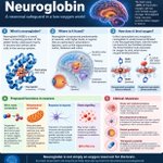
What if I told you that the brain has its own relative of haemoglobin, but its main job may not be to transport or store oxygen?
Meet neuroglobin, a neuronal protein that may help protect the brain not by supplying oxygen, but by limiting the biochemical damage caused by its absence. Surviving oxygen deprivation involves much more than oxygen delivery.

Rafael Sirera retweeted

“Expression of neuroglobin increases during hypoxia and is associated with greater neuronal survival.”
Fantastic pictorial of the biochemistry.

What if I told you that the brain has its own relative of haemoglobin, but its main job may not be to transport or store oxygen?
Meet neuroglobin, a neuronal protein that may help protect the brain not by supplying oxygen, but by limiting the biochemical damage caused by its absence. Surviving oxygen deprivation involves much more than oxygen delivery.
Rafael Sirera retweeted

I took about 50,000 photos through a 14" telescope last night to produce this: A hyper-detailed photo of the full moon. The fidelity of the photo was great enough to allow me to greatly enhance the color, revealing a hidden world of geological diversity.

Rafael Sirera retweeted
Do you know why hyperventilation can make you feel dizzy?
The answer lies in how the brain regulates its own blood supply.
⭐️ Like the kidneys, the brain must maintain relatively constant blood flow despite fluctuations in systemic arterial pressure.
1⃣ This is achieved through cerebral autoregulation, which adjusts the diameter of small arteries and arterioles to control vascular resistance.
➡️ When blood pressure falls, cerebral arterioles dilate, reducing resistance and helping preserve blood flow.
➡️ When pressure rises, they constrict, protecting delicate cerebral capillaries from excessive pressure.
2⃣ But the brain has another remarkable regulatory mechanism: its extraordinary sensitivity to carbon dioxide (CO₂).
CO₂ readily crosses the blood–brain barrier. An increase in CO₂ raises local H⁺ concentrations, promoting cerebral vasodilation and increasing blood flow. Conversely, when CO₂ levels fall, cerebral vessels constrict.
⭐️ And this brings us back to hyperventilation.
When you breathe excessively, you eliminate CO₂ faster than your body produces it. The resulting hypocapnia produces cerebral vasoconstriction, reducing blood flow to the brain.
➡️ The consequence? Dizziness, light-headedness and, in extreme cases, fainting — even when blood oxygen saturation remains perfectly normal.
However, cerebral autoregulation has limits. Severe hypotension can overwhelm compensatory vasodilation, leading to cerebral ischaemia, whereas severe hypertension can overcome protective vasoconstriction, potentially causing blood–brain barrier disruption and oedema.
The fascinating lesson is that delivering oxygen to the brain is not simply about how much oxygen is in your blood. It is also about how much blood actually reaches your neurons.
And sometimes, breathing more can paradoxically mean delivering less.

Rafael Sirera retweeted

¿Sabías dónde termina el agua de cada río de España y Portugal? 🌊🗺️
Este increíble mapa animado nos muestra el viaje a la inversa de nuestros ríos, desde su desembocadura en el mar hasta sus nacimientos en las montañas.
Créditos: u/depredador93
Do you know why hyperventilation can make you feel dizzy?
The answer lies in how the brain regulates its own blood supply.
⭐️ Like the kidneys, the brain must maintain relatively constant blood flow despite fluctuations in systemic arterial pressure.
1⃣ This is achieved through cerebral autoregulation, which adjusts the diameter of small arteries and arterioles to control vascular resistance.
➡️ When blood pressure falls, cerebral arterioles dilate, reducing resistance and helping preserve blood flow.
➡️ When pressure rises, they constrict, protecting delicate cerebral capillaries from excessive pressure.
2⃣ But the brain has another remarkable regulatory mechanism: its extraordinary sensitivity to carbon dioxide (CO₂).
CO₂ readily crosses the blood–brain barrier. An increase in CO₂ raises local H⁺ concentrations, promoting cerebral vasodilation and increasing blood flow. Conversely, when CO₂ levels fall, cerebral vessels constrict.
⭐️ And this brings us back to hyperventilation.
When you breathe excessively, you eliminate CO₂ faster than your body produces it. The resulting hypocapnia produces cerebral vasoconstriction, reducing blood flow to the brain.
➡️ The consequence? Dizziness, light-headedness and, in extreme cases, fainting — even when blood oxygen saturation remains perfectly normal.
However, cerebral autoregulation has limits. Severe hypotension can overwhelm compensatory vasodilation, leading to cerebral ischaemia, whereas severe hypertension can overcome protective vasoconstriction, potentially causing blood–brain barrier disruption and oedema.
The fascinating lesson is that delivering oxygen to the brain is not simply about how much oxygen is in your blood. It is also about how much blood actually reaches your neurons.
And sometimes, breathing more can paradoxically mean delivering less.
Rafael Sirera retweeted

EL MEJOR VIDEO DEL MES … LIBERACIÓN DE LA PUERTA DEL SOL ☀️.. UNA OBRA DE ARTE 👏👏👏


Rafael Sirera retweeted
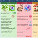
The other day, I was listening to @cienciabrujula on
@BrujulaOndaCero, where he was explaining what distinguishes animal cells from those of other eukaryotes. It inspired me to put together this infographic.
Plants, animals and fungi share the same fundamental eukaryotic cellular architecture, yet evolution has equipped them with remarkably different biochemical strategies.
Rafael Sirera retweeted

Bona vesprada, amics.
Finalment, hui vos porte el restant mapa del Regne de València i les seves jurisdiccions feudals.
Hui, la Governació de València.
Espere que vos agrade.

Rafael Sirera retweeted
Every drop counts: this diagram shows how water flow is ingested and secreted and later absorbed in the digestive system

Every drop counts: this diagram shows how water flow is ingested and secreted and later absorbed in the digestive system


Rafael Sirera retweeted
You have probably heard someone say, “I'm allergic to the cold!” But how is that possible? Cold, heat, sunlight and mechanical vibrations are physical stimuli, not allergens!
There is a group of conditions known as chronic inducible urticarias, in which physical stimuli trigger the activation of mast cells, causing the release of histamine and other inflammatory mediators.
Hence, mast cells do not always need IgE to become activated!
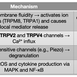
One potential mechanism involves TRP (Transient Receptor Potential) channels, a family of membrane proteins that act as molecular sensors for temperature, chemical signals and certain mechanical stimuli.
These channels are non-selective cation channels, permeable mainly to Ca²⁺ and Na⁺. When activated, they allow cations to enter the cell, altering intracellular signalling. In mast cells, increased cytosolic Ca²⁺ can contribute to degranulation and the release of histamine, leading to itching, redness and wheals.
However, TRP channels are only part of the story. Different forms of physical urticaria involve different molecular mechanisms, and some can indeed involve IgE-dependent mast cell activation.
Take home message: mast-cell activation is not synonymous with IgE-mediated allergy, and TRP channels represent one interesting potential route among several.
