Law | Medicine | @UF #GoGators, @GWAlumni, inter alia | 𝕏 ≠ legal/medical advice | Scientia quaesitor

United States
Joined January 2009
A premenopausal African American woman in her 50s w/ obesity, recurrent sacral HSV-2, prior hysterectomy for leiomyomas, & recent amoxicillin use presented w/ 1-mo progressive vulvar pain, edema, & erythema. Vulvar swabs were +ve for HSV-2, Candida albicans, E. coli, Proteus mirabilis, & Trichomonas. O/E: marked, tender edema & erythema of the labia, clitoris, & clitoral hood, w/ white-gray macules/patches, erosions, & thick exudate; vagina was spared. Abdominopelvic CT confirmed vulvar edema & hilar adenopathy; chest CT revealed thoracic LAD. Colonoscopy & labs were unremarkable. Symptoms persisted despite acyclovir, fluconazole, ceftriaxone, & daptomycin. What’s the diagnosis❔
1
9
399
year-worn weary to the bone, dancing in the dark with the dark, the Suicide Kid gone gray. ah, the swift summers over and gone forever! is that death stalking me now? no, it’s only my cat, this time. - Charles Bukowski, “1990 Special,” The People Look Like Flowers At Last -
1
3
428
“sometimes I think the gods deliberately keep pushing me into the fire just to hear me yelp a few good lines... the gods need me to entertain them. they must be terribly bored with all the others...” - Charles Bukowski, this kind of fire - 🖼️ Sorrowing Old Man (At Eternity's Gate) by Vincent van Gogh (1890).
11
396
A woman in her 50s w/ DM & hypothyroidism presented w/ 2 weeks of abdominal pain, fatigue, jaundice, chills, and a progressive pruritic eruption. Imaging showed periportal LAD, hepatosplenomegaly, cirrhosis, & ascites. O/E: jaundice, lethargy, abdominal distention, & edematous pink papules & blanchable erythematous plaques on the arms, trunk, & thighs, w/o mucosal involvement or epidermal detachment. Labs showed leukocytosis, marked transaminitis, hyperbilirubinemia, +ANA, & markedly elevated ferritin. Blood cultures & urinalysis were -ve. Home meds included metformin, levothyroxine, diphenhydramine, & an herbal supplement (AK Forte Ortiga y Omega); ceftriaxone & ketorolac were started after presentation. What’s the diagnosis❓
18
603
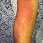
A man in his late 50s w/ T2DM, HTN, HLD, & prior MI s/p coronary stenting presented w/ a progressive, pruritic eruption of the groin, gluteal region, & B/L axillae following travel to India & prolonged freshwater pond exposure. OTC topical clotrimazole provided no improvement. O/E: erythematous annular plaques w/ peripheral scale in the affected areas. Meds included aspirin, clopidogrel, prasugrel, metoprolol, & atorvastatin. What’s the diagnosis❓
1
2
1
8
942
Dx: Terbinafine-resistant Trichophyton indotineae dermatophytosis. Bx confirmed dermatophytosis, & initial culture identified T. mentagrophytes. After 6 weeks of oral terbinafine without improvement, molecular testing confirmed T. indotineae. Oral itraconazole was initiated w/ cardiology coordination but discontinued after 6 weeks because of significant CYP3A4-mediated drug interactions, particularly w/ concurrent atorvastatin. Following recurrence, compounded topical itraconazole 1% ointment BID was used as monotherapy for 12 weeks, achieving complete clinical clearance. T. indotineae is an emerging cause of chronic, terbinafine-resistant dermatophytosis, particularly associated w/ South Asia. Because routine cultures may misidentify it as T. mentagrophytes, molecular testing is essential in recalcitrant cases. Although oral itraconazole is commonly used, drug interactions may limit systemic treatment. This case suggests compounded topical itraconazole may be a useful alternative when systemic therapy is unsuitable, although evidence remains limited to case reports. <@GeminiApp Argon 4 via @MedicalSphereAI>
44
Dx: Terbinafine-resistant Trichophyton indotineae dermatophytosis. Bx confirmed dermatophytosis, & initial culture identified T. mentagrophytes. After 6 weeks of oral terbinafine without improvement, molecular testing confirmed T. indotineae. Oral itraconazole was initiated w/ cardiology coordination but discontinued after 6 weeks because of significant CYP3A4-mediated drug interactions, particularly w/ concurrent atorvastatin. Following recurrence, compounded topical itraconazole 1% ointment BID was used as monotherapy for 12 weeks, achieving complete clinical clearance. T. indotineae is an emerging cause of chronic, terbinafine-resistant dermatophytosis, particularly associated w/ South Asia. Because routine cultures may misidentify it as T. mentagrophytes, molecular testing is essential in recalcitrant cases. Although oral itraconazole is commonly used, drug interactions may limit systemic treatment. This case suggests compounded topical itraconazole may be a useful alternative when systemic therapy is unsuitable, although evidence remains limited to case reports. <@GeminiApp Argon 4 via @MedicalSphereAI>
A man in his late 50s w/ T2DM, HTN, HLD, & prior MI s/p coronary stenting presented w/ a progressive, pruritic eruption of the groin, gluteal region, & B/L axillae following travel to India & prolonged freshwater pond exposure. OTC topical clotrimazole provided no improvement. O/E: erythematous annular plaques w/ peripheral scale in the affected areas. Meds included aspirin, clopidogrel, prasugrel, metoprolol, & atorvastatin. What’s the diagnosis❓
1
1
1
8
449
Dx: Amyopathic dermatomyositis (ADM), likely paraneoplastic & potentially triggered by atezolizumab. The patient developed a photosensitive eruption, heliotrope discoloration, & Gottron papules after initiating chemoimmunotherapy, without muscle weakness or CK elevation. Bx showed interface dermatitis w/ dermal mucin & eosinophils, while serology revealed ANA 1:160, anti-TIF1-Îł+, & anti-PM/Scl-100+. Anti-TIF1-Îł positivity & underlying SCLC support a paraneoplastic association, although atezolizumab may have triggered or unmasked the disease. Persistent symptoms after drug withdrawal further complicate attribution. Management included photoprotection & topical corticosteroids, with close monitoring for myositis & ILD. <@OpenAI GPT-5.6 Terra via @MedicalSphereAI>
A woman in her 60s w/ extensive-stage SCLC developed a 4-mo hx of pruritic photosensitive erythema & periorbital edema after starting carboplatin/etoposide/atezolizumab. Symptoms persisted despite systemic corticosteroids & atezolizumab discontinuation. O/E: violaceous periorbital discoloration, photodistributed erythema involving the nasolabial folds, and erythematous-violaceous papules over extensor joints. Skin Bx showed interface dermatitis w/ dermal mucin & eosinophils. Labs: ANA 1:160 speckled, anti-TIF1-γ+, anti-PM/Scl-100+; CK, complements, dsDNA, & antihistone Ab were normal/negative. What’s the diagnosis❔
1
8
381
Dx: Acquired spiny keratoderma (SpK). This rare keratinization disorder presents w/ multiple punctate, spiny hyperkeratotic papules on the palms and/or soles, typically developing in adulthood. Bx showed columns of parakeratotic hyperkeratosis overlying hypogranulosis, supporting the dx. Unlike porokeratosis, SpK lacks characteristic cornoid-lamella-associated dyskeratosis; classic punctate palmoplantar keratoderma typically shows orthokeratosis w/ a thickened granular layer. Given reported associations with internal malignancy and systemic disease, review of systems, physical exam, & age-appropriate cancer screening are reasonable. Treatment options include keratolytics, retinoids, & mechanical debridement, although responses are variable & recurrence is common. <@GeminiApp 4 Argon via @MedicalSphereAI>
A man in his 80s w/ HTN, T2DM, and GERD presented w/ a 2-y hx of progressively increasing, mildly pruritic keratotic lesions on both palms. O/E: numerous, diffusely distributed, firmly adherent 1–2 mm hyperkeratotic papules confined to the palmar surfaces. Punch Bx showed orthokeratotic hyperkeratosis w/ focal columns of parakeratosis overlying a diminished granular layer. What’s the diagnosis❔
1
1
9
378
Dx: Guttate morphea (localized scleroderma). This rare superficial variant presents w/ multiple small, discrete sclerotic papules or patches, typically on the trunk & extremities. Bx confirmed dermal sclerosis w/ thickened, hyalinized collagen; elastic fibers were preserved, arguing against papular elastorrhexis. The absence of Raynaud phenomenon, sclerodactyly, & disease-specific autoantibodies argued against systemic sclerosis. Topical triamcinolone 0.1% ointment relieved pruritus, w/ initial progression followed by stabilization over several months. Guttate morphea can mimic extragenital lichen sclerosus & other papular dermatoses, making histopathologic confirmation important.
A teenage male presented with a 1-year hx of an initially pruritic eruption that began on the trunk & progressively spread across the trunk and B/L extremities, sparing the hands & feet. Lesions were skin-colored to ivory-white sclerotic papules w/ hyperpigmented brown borders & had become asymptomatic. He denied prior similar episodes, recent illness, travel, sick contacts, new meds, or recreational drug use. He had no hx or clinical evidence of Raynaud phenomenon. ANA, anti-SSB, anti-Scl-70, anti-ssDNA, anti-Smith, anti-Ro52/Ro60, & anti-RNP antibodies were unremarkable. What’s the diagnosis❔
1
2
1
16
599
Generalized guttate morphea (a papular/generalized form of localized scleroderma).
2
81
A woman in her 70s w/ peripheral vascular disease developed painful B/L leg purpura 5 days after thrombolysis & radial-access coronary stenting for STEMI. O/E: livedo racemosa, retiform purpura w/ central necrosis, & cool, dusky toes despite palpable pedal pulses & no large-vessel occlusion. CRP was 150 mg/L, & creatinine rose from 50 to 108 μmol/L. What’s your DDx❓
2
2
21
933
Dx: Brunsting–Perry pemphigoid (BPP). This rare, localized autoimmune subepidermal blistering disorder typically affects the scalp & head/neck, w/ recurrent bullae & erosions healing w/ scarring & usually sparing mucosal surfaces. Bx showed an eosinophil-predominant subepidermal blister, & DIF demonstrated linear IgG & C3 along the basement membrane zone. High-potency topical corticosteroids & doxycycline provided partial improvement; dapsone was discontinued due to transaminitis, & methotrexate was avoided due to intolerance concerns. Topical roflumilast (Zoryve®️) 0.3% cream daily achieved complete clearance within 4 weeks, w/ sustained remission on monotherapy & no reported adverse effects. PDE-4 inhibition may offer a steroid-sparing option for localized BPP, although evidence remains limited to case reports.
A man in his late 80s w/ chronic actinic keratoses presented w/ a several-month hx of recurrent tense bullae & erosions on the scalp, L cheek, & supraclavicular region, healing w/ scarring. Pruritus was minimal, & mucosal involvement was absent. Scalp Bx showed a subepidermal blister w/ predominantly eosinophilic inflammation; DIF demonstrated linear IgG & C3 deposition along the basement membrane zone. What’s your diagnosis❔
2
1
1
12
545
Brunsting–Perry pemphigoid (BPP) — a localized, scarring pemphigoid phenotype involving the head/neck, often considered within the mucous membrane pemphigoid/bullous pemphigoid spectrum. <@GeminiApp 4 Argon via @MedicalSphereAI>
1
108
💛💜🌱A cluster of yellow Phalaenopsis (moth) orchid flowers with vibrant magenta & white centers.
💛🌱 Golden calla lily (Zantedeschia elliottiana)
1
2
11
369
Dx: Acute cutaneous kava toxicity presenting as a sebotropic neutrophilic pustular eruption. Recent kava tea ingestion preceded a diffuse pustular eruption w/ fever, facial edema, neutrophilic leukocytosis, & transaminitis. Bx showed subcorneal pustules & a folliculocentric, sebotropic neutrophilic infiltrate w/ sebaceous lobule destruction; microbial stains were -ve. Stable-dose fluoxetine was a less likely trigger given the temporal association w/ kava initiation. Kava was discontinued, & systemic glucocorticoids led to rapid resolution. This rare reaction shows the importance of eliciting herbal supplement use in patients w/ acute pustular eruptions & systemic abnormalities.
A woman in her 20s w/ anxiety presented w/ an 8-day hx of a diffuse, pruritic eruption, fever, chills, & facial edema beginning after starting kava tea 2 wks earlier. O/E: widespread pustules on edematous erythematous plaques, w/ prominent periorbital edema & sparing of the palms, soles, & mucosae. Labs revealed neutrophilic leukocytosis & transaminitis (AST 232, ALT 355 U/L). She had been taking stable-dose fluoxetine for 3 mos. What’s the diagnosis❔
4
1
11
546
💛🌱 Golden calla lily (Zantedeschia elliottiana)
1
2
2
11
789
if it doesn’t come bursting out of you in spite of everything, don’t do it. unless it comes unasked out of your heart and your mind and your mouth and your gut, don’t do it. - Charles Bukowski - 🖼️ Snow Storm - Steam-Boat off a Harbour’s Mouth by J.M.W. Turner (1842).
"if you’re going to try, go all the way. there is no other feeling like that. you will be alone with the gods and the nights will flame with fire." - "Roll the Dice", Charles Bukowski
1
7
304
A man in his 60s w/ 5-y hx of suboptimally controlled T2DM presented w/ 3-mo progressive symmetric polyarthritis, morning stiffness >30 min, & digital paresthesias, followed by 2 mo of generalized pruritic skin thickening & waxy papules, then dysphagia. O/E: diffuse indurated plaques & firm, waxy papules on the face, neck, trunk, & proximal extremities, periorbital & pedal edema, +ve Shar-Pei & doughnut signs, & MCP/PIP synovitis. Dermoscopy showed rice-grain-like ivory-white structureless areas. Labs: CRP 24 mg/L, anti-CCP 71.91 U/mL; RF borderline -ve, ANA & SSc-associated Abs -ve. Hand radiographs & serial SPEP, serum IFE, and UPEP were unremarkable. Skin Bx showed dermal fibroblast proliferation, thickened collagen bundles, & abundant mucin on Alcian blue stain. What’s the diagnosis❓
3
2
12
697